
Figure 6. Sleep-deprived flies have reduced MIP levels in neurons innervating the optic lobe medulla.
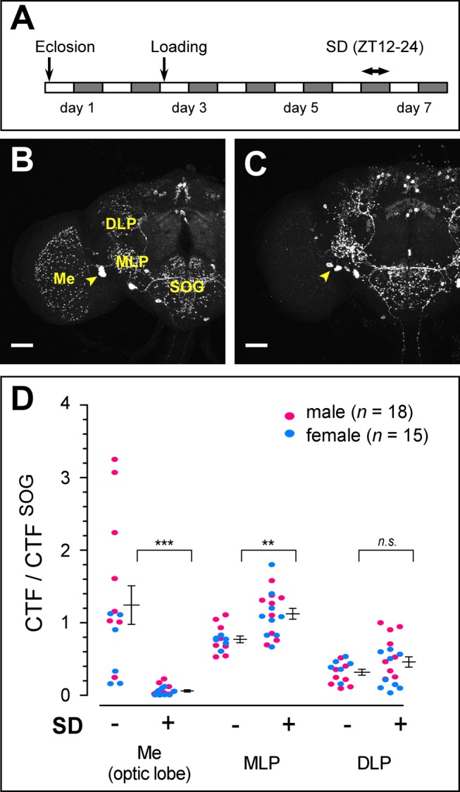
(A) Protocol for sleep deprivation (SD). (B, C) Representative confocal images of anti-MIP staining of the w1118 female brain subjected to non-SD (B) and SD (C) conditions. Yellow arrowheads indicate somata of MIP-LMIo. (D) Normalized MIP-immunoreactivity of non-SD (−) versus SD (+) female and male flies in the indicated areas of interest. Data are shown as means ± SEM. **, p<0.01, ***, p<0.001 for the comparison by Student's t test. Scale bars, 50 µm. Me, medulla; DLP, dorso-lateral protocerebrum.