
Figure 8. Axonal processes of MIP-LMIo neurons innervate the dendritic field of pdf neurons.
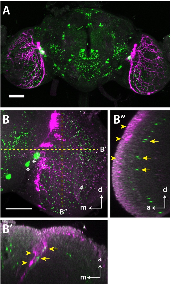
(A) A confocal image of pdf-Gal4/UAS-EGFP fly brain stained with anti-MIP (green) and anti-EGFP (magenta) antibodies. The brain is oriented with dorsal up. (B) A high magnification confocal image of the optic lobe of pdf-Gal4/UAS-DenMark fly brain stained with anti-MIP (green) and anti-RFP (magenta) antibodies. Anti-RFP labels a dendrite marker, DenMark, which visualizes the dendritic field of pdf neurons. Asterisks indicate somata of MIP-LMIo neurons. Sections perpendicular to the dotted yellow lines are shown separately in (B′) and (B″). (B′) shows MIP-immunoreactive processes (yellow arrows) innervate the dendritic field of pdf neurons (yellow arrowheads). (B″) shows some MIP-labeling (arrows) occurs near the dendritic field of pdf neurons (arrowheads), indicating that MIP can also function as a paracrine factor. White arrows labeled with d, a, m indicate dorsal, anterior, and medial orientations, respectively. Scale bars, 50 µm.