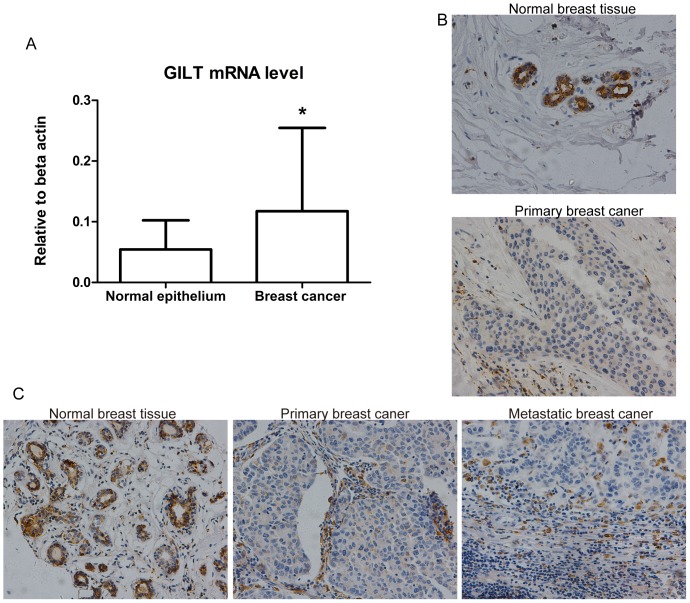
Figure 3. Involvement of GILT in tumorigenesis and lymph node metastasis in breast cancer.
(A) GILT mRNA expression increased in malignant cells compared with adjacent normal epithelial cells. The real-time PCR results confirmed that there was a 2.18-fold up-regulation of GILT mRNA in breast cancer cells compared with adjacent normal epithelial cells (* P<0.05, n = 19). Relative means ± standard deviation for GILT mRNA obtained from tumor tissue and normal adjacent tissue are shown. (B) GILT protein expression decreased in malignant tissues compared with adjacent normal epithelial tissues. The immunohistochemistry results confirmed that 78.95% (15/19) showed weaker staining in carcinoma tissue than in adjacent normal breast tissue; representative images are shown (original magnification, ×200). (C) GILT protein expression in matched normal-cancerous-metastatic breast tissues (n = 44). Representative GILT expression in respective normal-cancerous-metastatic breast tissues sections from one patient. Both intensity and proportion score of GILT staining in primary breast cancer as well as metastatic cancer tissue were 0, compared with 2 and 4 respectively in normal breast tissue (original magnification, ×200).

