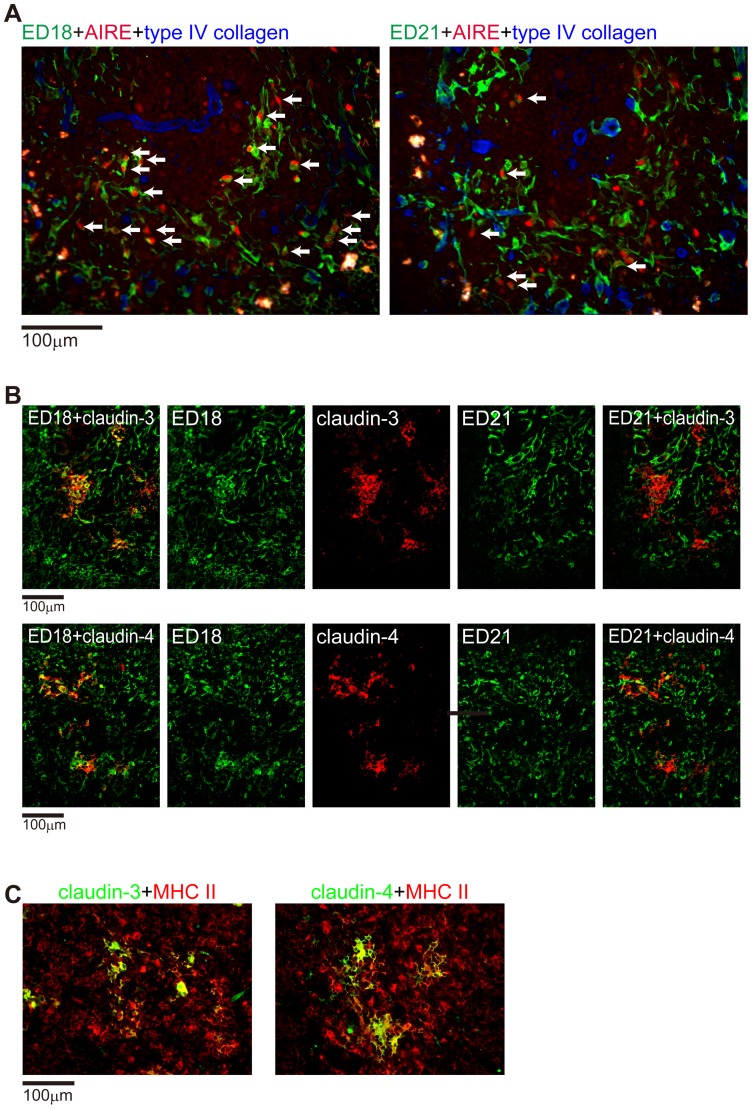
Figure 6. Expression of functional molecules in rat mTEC1 and mTEC2 subsets.
(A) Rat thymic sections were stained with Alexa488-conjugated ED18 or ED21 (green) and anti-AIRE antibody followed by Alexa594-conjugated anti-goat IgG (red). Tissue framework was stained blue with anti-type IV collagen followed by AMCA-conjugated anti-rabbit IgG. Arrows indicate AIRE expression associated with ED18- and ED21-positive cells. (B, C) Rat thymic sections were stained with anti-claudin-3 or anti-claudin-4 antibodies followed by AMCA-conjugated anti-rabbit IgG, Alexa488-conjugated ED18, Alexa594-conjugated ED21, and Alexa647-conjugated anti-rat MHCII. Pseudocolors were assigned using AxioVision software.

