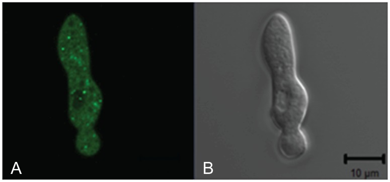
Figure 6. PALA is localized to the small intracellular vesicles.
GFP-tagged PALA was expressed under the regulation of the ccg-1 promoter in a ΔpalA isolate. The GFP fluorescent image of a germling is shown in the panel on the left (A). The DIC image of the same germling is shown in the right panel (B). The bar in the DIC image is 10 µm in length.

