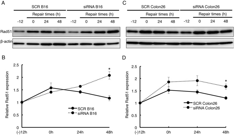
Figure 6. Prolonged Rad51 expression after DNA damage by etpoposide in cancer cells.
SCR or SLD5 siRNA transfected B16 (A, B) or Colon26 (C, D) cells were treated with etoposide for 12 h (−12∼0). Cells were lysed at the indicated times. Western blot analyses of Rad51 expression is shown. β-actin was the internal control. Data were quantitatively evaluated based on densitometric analysis. Results are fold-change compared with the level seen in SCR B16 (B) or SCR Colon26 (D) before treatment with etoposide. Data represent the mean ± SD.*, P<0.05 (n = 3).

