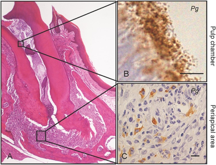
Figure 1. Dental Pg infection induces periapical granuloma.
Severe pulp necrosis is observed in the first upper molars after six weeks of Pg infection. (A) Periapical granuloma is a representative histological feature of Pg infected molars, and is infiltrated with neutrophils and macrophages. (B) Immunohistochemical staining shows Pg colonies (brown pigment) in the pulp chambers and (C) in neutrophils and macrophages in the periapical area. Scale bar, 10 µm. Pg, Porphyromonas gingivalis. Experiments were performed three times or more with similar results.

