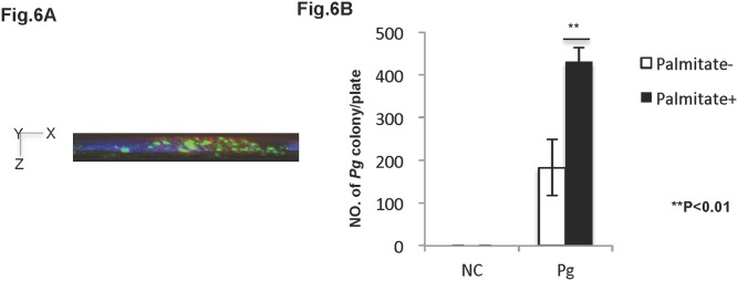
Figure 6. Palmitate increases Pg cell invasion in HuhT1 cells.

Antibody protection assay (see Material & Methods) was used to determine Pg invasion in HuhT1 cells. (A) Invaded Pg was analyzed by fluorescent microscopy at magnification of 1000x. Side view (X–Z plane) is shown. Pg (green); DAPI (blue); Phalloidin (red). (B) Pg invasion into HuhT1 cells is increased under palmitate pre-treatment. Mean ± SD, **P<0.01. NC, negative control; Pg Porphyromonas gingivalis. Experiments were performed three times or more with similar results.
