
Figure 5.
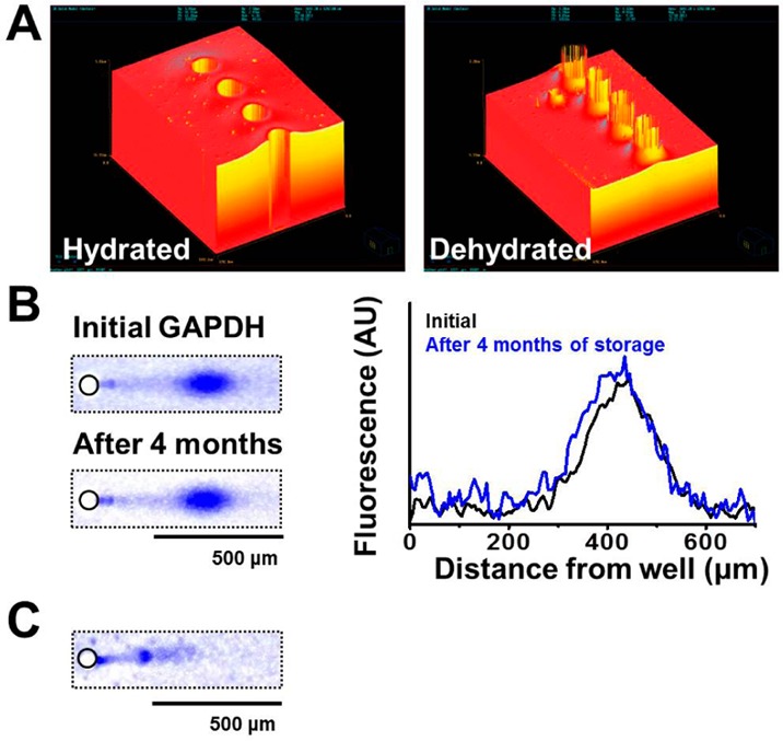
Archival storage of scWestern slides with subsequent successful antibody probing. (A) Optical profilometer images of hydrated and dehydrated scWestern PA gel features. Circular features are microwells, 30 μm in diameter. (B) False-color fluorescence micrographs and intensity traces of the same scWestern device showing antibody probing for GAPDH (blue signal) after fresh preparation and after 4 months of archival storage. (C) Fluorescence micrographs of the same scWestern device reprobed for β-tubulin (blue signal). AU, arbitrary unit. E = 40 V cm–1, lysis time = 25 s, electrophoresis time = 20 s, 12%T PA gel (GAPDH, Alexa Fluor 647-labeled secondary antibody; β-tubulin, Alexa Fluor 594-labeled secondary antibody).