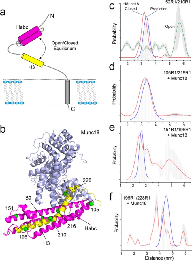
Figure 4.

Conformational heterogeneity in the neuronal SNARE protein syntaxin 1A. (a) Syntaxin 1A is believed to exist in an open–closed equilibrium, where the H3 motif (yellow) can be either associated with or dissociated from the regulatory Habc domain (magenta). (b) The crystal structure of the syntaxin 1A/munc18-1 complex (PDB ID 3C98), where syntaxin is in a closed conformation and the H3 motif is resolved and folded along the Habc domain; pairs of labels were placed across syntaxin to make distance measurements between the H3 and Habc regions and along the H3 motif.40 (c) Distance distributions obtained for 52R1/210R1 in full length membrane reconstituted syntaxin in the absence (green trace) and presence of munc18 (red trace); the blue trace shows the prediction based upon the munc18/syntaxin crystal structure. (d, e, f) distance distributions obtained in the presence of munc18 for syntaxin(1–262) for 105R1/216R1, 151R1/196R1, and 196R1/228R1, respectively; the blue trace shows the distance distribution predicted based upon the munc18/syntaxin crystal structure.
