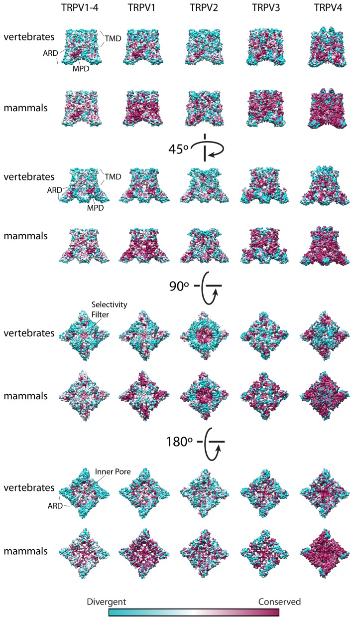
Figure 3. Tridimensional Conservation plots for TRPV1-4 comparing vertebrate and mammalian sequences.
Conservation degree for each amino acid position was plotted on the solved structure for TRPV1 (pdb code 3J5P) for the MSAs for TRPV1-4 and TRPV1. For the conservation plot of TRPV2, TRPV3 and TRPV4 homology models were built based on the coordinates of TRPV1 (pdb code 3J5P). The conservation ranges from cyan (divergent) to magenta (conserved). Specific domains are indicated: TMD, transmembrane domain; ARD, ankyrind repeat domain; MPD, membrane proximal domain.

