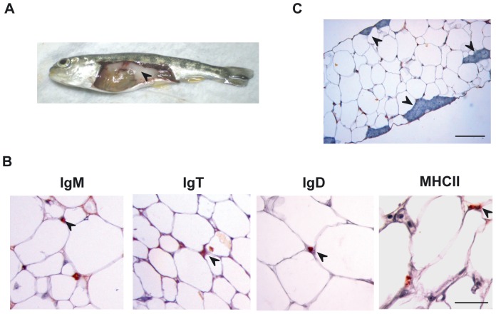
Figure 1. Immunohistological analysis of visceral rainbow trout AT.
(A) The rainbow trout visceral AT (black arrow) was removed, fixed in Bouin's solution, embedded in paraffin and sectioned at 5 µm. After dewaxing and rehydration, sections were subjected to an indirect immunocytochemical method to detect trout IgM, IgT, IgD and MHC-II (B) Arrow heads point to representative positive staining. Scale bars, 50 µm. (C) Representative photomicrograph of an IgM immunostained section showing structures that resemble mammalian milky spots (arrow heads). Scale bars, 100 µm.

