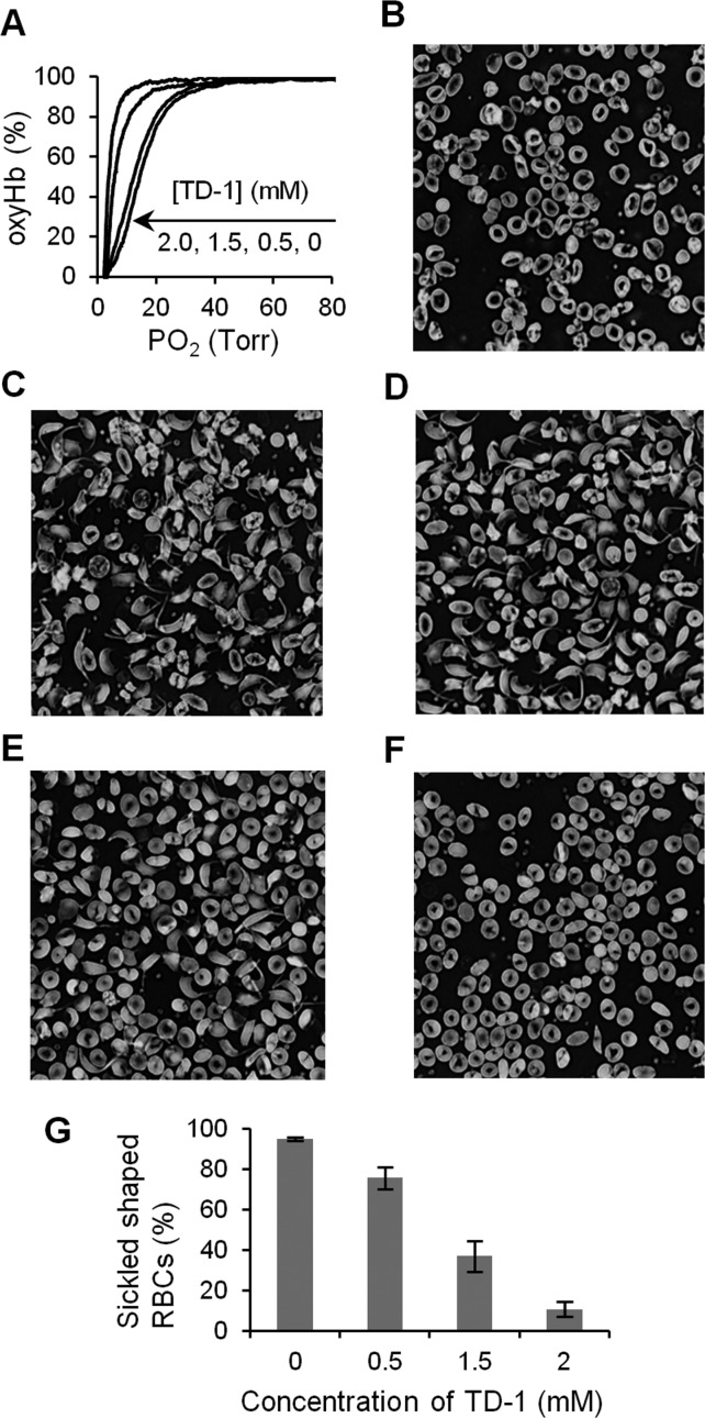
Figure 4.
Anti-sickling effect of TD-1. (A) Representative ODCs of the hemolysates of SS RBCs without or with TD-1 at 25 °C. The ODC shifted to the left in a dose-dependent manner of TD-1. P50 of hemolysates from SS RBCs (hematocrit ∼20%) treated without or with 0.5, 1.5, and 2.0 mM of TD-1 were 13 ± 0.5 (data mean value ± s.d.), 12 ± 0.8, 7.2 ± 0.5, and 5.1 ± 1.1 Torr (P < 0.001, vs without TD-1), respectively. (B) Morphology of SS RBCs (hematocrit ∼20%) incubated under normoxic conditions revealed primarily discocytes with some irreversibly sickled cells. (C) Morphology of SS RBCs incubated with 4% oxygen at 37 °C for 3 hours revealed sickling of RBCs. SS RBCs were mixed with 0.5 mM (D), 1.5 mM (E), and 2 mM (F) of TD-1 before incubation with 4% oxygen at 37 °C for 3 hours. (G) Treatment with TD-1 reduced the SS RBC sickling induced by hypoxia in a dose-dependent manner. Error bars represent standard deviation.

