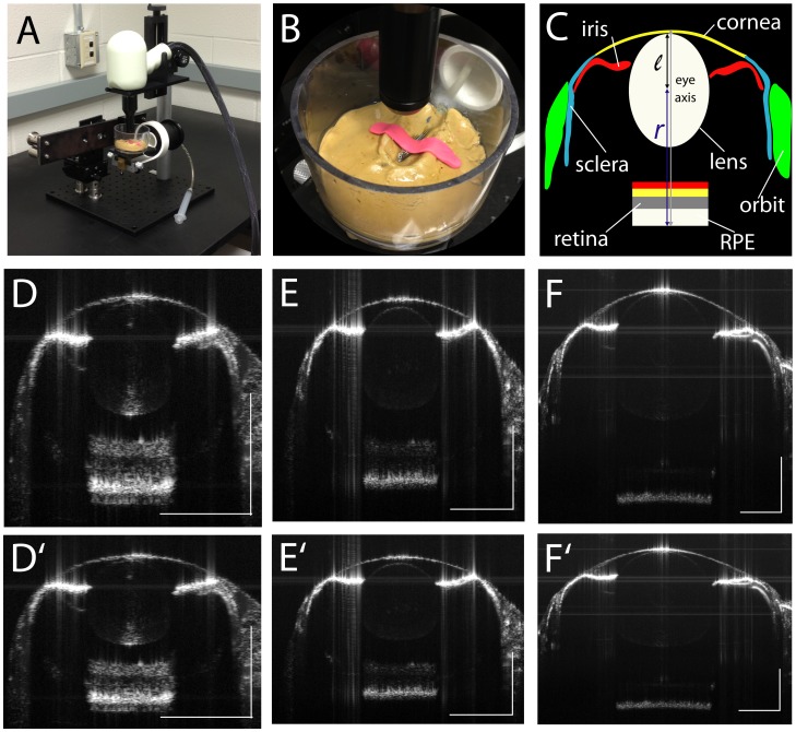
Figure 1. SD-OCT imaging stage and B-scan examples.
A. Imaging stage for Bioptigen Envisu 2200 with zebrafish immersion cuvette. B. Zebrafish immobilized using a strip of modeling clay to prevent movement or floating during immersion. C. Schematic showing highly reflective structures of the zebrafish eye traced over 1 mpf B-scan; l, lens radius; r, retinal radius. D. 15 dpf; E. 1 mpf; F. 2 mpf. Scale bars: 300 µm. D′, E′, F′: as above with aspect ratio corrected to 1∶1. As zebrafish eyes age and increase in size, the reflected signal from the retina is reduced, making lamination less visible, though the strongly hyper-reflective RPE can still be observed.

