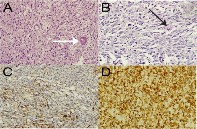
Figure 4 A–D:
Images of the histological specimen. Haematoxylin and eosin stains showed (A) a sheet of round to spindle-shaped malignant cells with an entrapped bile duct (white arrow) at ×20 magnification and (B) the typical presence of cytoplasmic eosinophilic hyaline globules (black arrow) at ×40 magnification. Immunohistochemistry showed positivity for (C) desmin and (D) myogenic differentiation antigen 1 at ×40 magnification.

