Figure 5.
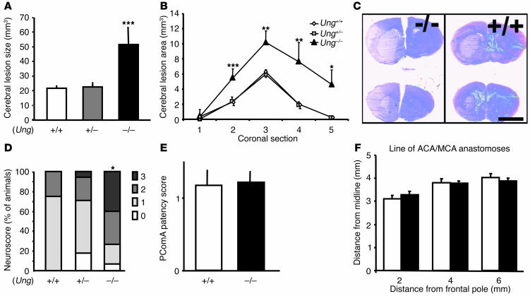
Effects of focal-brain ischemia in Ung–/–, Ung+/–, and Ung+/+ littermate mice. (A) Brain lesion volume and (B) lesion areas in Ung–/– mice compared with Ung+/– and Ung+/+ WT littermate mice after 30 minutes of MCAo and 72 hours of reperfusion. Brain lesion areas were determined on five anterior-posterior serial coronal H&E-stained cryostat sections (20 ∝m); mean ± SEM of 10 to 15 animals per group; **P < 0.01, ***P < 0.005, ANOVA and Tukey post hoc test. (C) Typical H&E-stained 20 ∝m brain sections from Ung–/– and Ung+/+ littermate mice. Scale bar: 3 mm. (D) Neurological sensory-motor deficits were determined after 72 hours and scored from 0 (no deficit) to 3 (severe); *P < 0.05, ANOVA on ranks (Kruskall Wallis). (E) The development of left and right posterior communicating arteries (PcomA) was determined in carbon black_perfused brains as follows: 0, absent; 1, present but poorly developed (hypoplastic); and 2, well formed; mean ± SEM from eight animals per group; ANOVA on ranks (Kruskall Wallis). (F) The distance from midline of the line of anastomoses between the ACA and the MCA was determined in carbon black_perfused brains; mean ± SEM from eight animals per groups. ANOVA and Tukey post hoc test.

