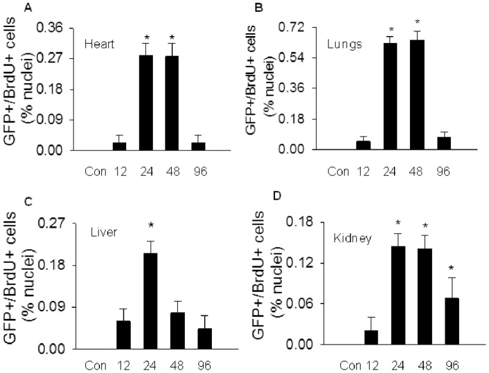
Figure 2. Characterization of BMDEPC proliferation in multiple organs.
WT-GFP-BM mice were injected with saline (Con) or LPS and BrdU (100 μg/kg, i.v., to label proliferating cells). Organs were harvested at 12, 24, 48 or 96 hours after LPS and 4 hours after BrdU injection. Cryosections were prepared and stained with GFP plus BrdU antibodies to identify proliferating BMDEPCs, and nuclei counterstained with DAPI. Number of GFP+/BrdU+ proliferating BMDEPCs on each slide from heart (A), lung (B), liver (C) and kidney (D) was counted and expressed as a percentage of total cells as revealed by DAPI nuclear staining. Mean ± SEM of 5 mice per group. *, p<0.05, compared with control group. There was an organ-dependent variation in numbers and kinetics of BMDEPC proliferation.

