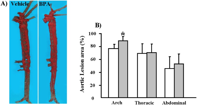
Figure 4. Quantitative analysis of aortic atherosclerosis in WHHL rabbits at 12 weeks.
(A) Representative micrographs of the aortic trees stained with Sudan IV. (B) The sudanophilic en-face lesion areas in different parts of aortic trees were determined using an image analysis system as described in the Materials and Methods section. Data are expressed as the means ± SD, n = 6 for each group. *p<0.05 vs. vehicle.

