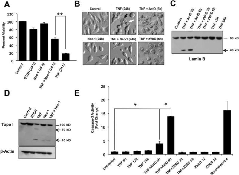
Figure 1. Induction of necroptosis by TNF-α in L929 cells.

A. MTT viability assays showing that treatment of L929 cells with 30 μM Nec-1 significantly attenuated TNF-α-induced cytotoxicity compared to treatment with TNF-α alone (**P < 0.01). Ethanol (ETOH, 1 mM) was used as vehicle control. B. Microscopic analysis of TNF-α-treated L929 cells showed necrosis-like morphologic features such as condensed nuclei and swelled cytoplasms, with no apoptotic blebbing, and partial restoration of normal cell morphology in the presence of Nec-1. The combination of TNF-α plus Actinomycin D (ActD) induced the classical apoptotic morphology characterized by cellular blebbing and apoptotic bodies, whereas the combination of TNF-α plus zVAD induced extensive necrosis. The black arrow points to a round swelled cell whereas the white arrow points to a small round cell with pyknotic nuclei and little cytoplasm. C. Immunoblots of lysates from L929 cells exposed to TNF-α/ActD showed cleavage of Lamin B into its signature 46 kD apoptotic fragment but no cleavage was observed in cells treated with TNF-α alone or TNF-α/zVAD. D. Immunoblots of lysates from L929 cells exposed to TNF-α alone also showed cleavage of Topo I into its necrotic signature fragments of 70 kD and 45 kD, with Nec-1 inhibiting this cleavage. β-actin was used as loading control. E. Caspase-3 activity, measured with the Ac-DEVD-AMC substrate, was detected in cells treated with TNF-α/ActD but not in cells treated with TNF-α alone or TNF-α/zVAD (*P < 0.05). Staurosporine, a classical inducer of apoptosis and caspase-3, served as positive control for caspase activation. The results shown in A and E are from at least three independent experiments performed in duplicates or triplicates.
