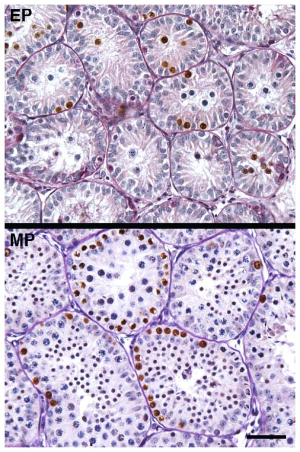
Figure 4.
Profiles of BrdU labelled nuclei (brown) in the testis of an early pubertal (EP) and mid pubertal (MP) rhesus monkey. Note the absence of BrdU label in the interstitium, which is in striking contrast to robust S-phase labelling of cells within the seminiferous tubules. The absence of BrdU labeling in the interstitium from testes of EP and MP monkeys may be compared to the robust HSD3B signal observed at these stages of development and shown in Figure 2. Scale bar, 50 μm. For photomicrographs illustrating the typical architecture of the testis under control conditions and the stimulation paradigm the reader is referred to Ramaswamy et al 2000.

