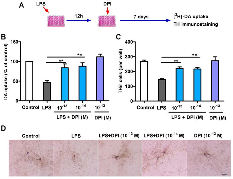
Figure 3.
Dopaminergic neuroprotection by post-treatment with subpicomolar DPI 12 h after inflammatory challenge in primary neuron-glia cultures. (A) Experimental designs. Midbrain neuron-glia cultures were pre-treated with LPS (20 ng/ml) for 12 h, followed by DPI (10−14 or 10− 13 M) treatment. (B) [3H]-DA uptake assay and (C) THir neuron count analysis revealed significant dopaminergic protection 7 days after DPI treatment. (D) Representative cell images of TH immunostaining 7 days after DPI treatment indicate prominent protection of the dopaminergic neuronal cell bodies and dendrites. The results of DA uptake are expressed as a percentage of the controls and are the mean ± SEM from three to four experiments performed in duplicate. The results of THir cell counts are expressed as the mean ± SEM from three to four experiments performed in duplicate. Data were analyzed using one-way ANOVA, followed by Bonferroni’s post hoc multiple comparison test. **p < 0.01; Bar = 50 μm.

