
Figure 1.
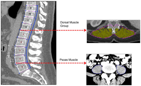
Preoperative CT imaging was used to measure psoas and dorsal muscle group size. After identifying individual vertebral levels, the imaging slices at the inferior aspects of L4 and T12 vertebrae were selected. The total cross-sectional area of the psoas muscles was measured at the L4 level; dorsal muscle group area was measured at the T12 vertebrae.