Figure 1.
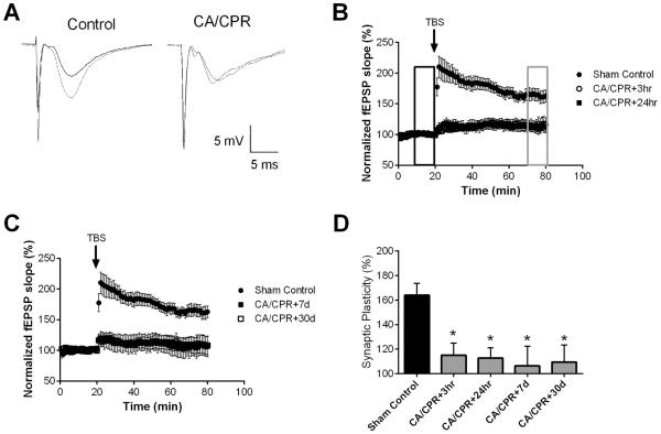
Ischemia impairs synaptic plasticity in the hippocampus. A) Example fEPSPs from sham operated control and 30 day after CA/CPR mice before (black) and after (grey) TBS. B) Time course of fEPSP slope (mean ± SEM) from sham (solid circles) and mice 3 hrs (open circles) or 24 hrs (solid squares) after cardiac arrest and cardiopulmonary resuscitation (CA/CPR). Arrow indicates timing of theta burst stimulus (TBS; 40 pulses). C) Time course of fEPSP slope (mean ± SEM) from sham (solid circles) and mice 7 days (open circles) or 30 days (solid squares) after cardiac arrest and cardiopulmonary resuscitation (CA/CPR). Arrow indicates timing of theta burst stimulus (TBS; 40 pulses) D) Quantification of change in fEPSP slope following TBS. Average fEPSP slope (mean ± SEM) 60 minutes after TBS (in grey box in B normalized to baseline (black box in B). * P < 0.05 compared to sham controls.

