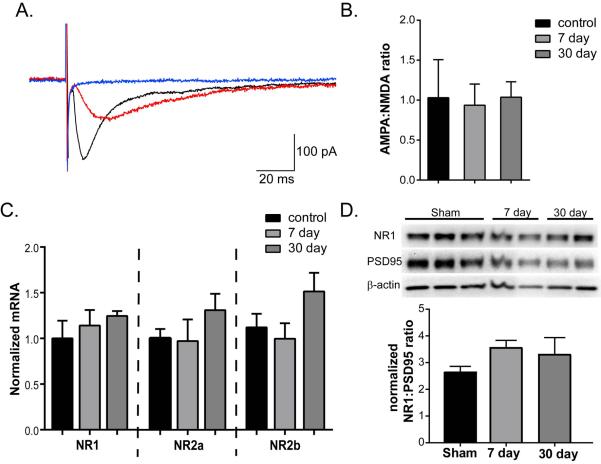
Figure 4.
Ischemia does not alter AMPA/NMDA ratio. A) Representative EPSCs from sham control. Application of NBQX (red trace) inhibits the AMPA component of the initial EPSC (black trace). Subsequent application of D-APV inhibits to remaining NMDA component (blue trace). B) Quantification of AMPA to NMDA ratio for all cells recorded demonstrates no difference of CA/CPR. C) Quantification of mRNA expression of the 3 predominant isoforms of NMDA receptor expressed in the hippocampus, GluN1, 2A and 2B. D) Representative Western blot analysis of NR1, PSD95 and β-actin in synaptic membrane preparations from hippocampi obtained at either 7 or 30 days after CA/CPR, or sham control mice. Quantification (bottom) of NR1:PSD95 ratio shows not changes in response to CA/CPR. Data presented as mean ± SEM.

