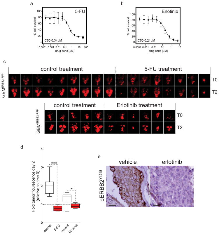
Figure 4. Zebrafish brain tumor models can be used for preclinical drug testing.
(a) Single cell suspensions of 750 GBMERBB2-RFP tumor cells were seeded into each well of a 96-well plate in neurobasal medium exactly as described7. 24 hours later cells were dosed with the indicated concentrations of (a) 5-FU or (b) Erlotinib, or DMSO vehicle control and incubated for a further 72 hours. Percent cell survival relative to DMSO only controls was then determined in each well using the Cell Titer Glo reagent (Promega) and Envision plate reader (Perkin-Elmer). Assays were performed in independent triplicates. (c) Zebrafish harboring GBMERBB2-RFP tumors were established exactly as described in Figure 1. After 24 hours zebrafish were treated with addition of 5-FU or vehicle (control) to the tank water (top), or vehicle (control) or Erlotinib by oral gavage on two consecutive days. All zebrafish were imaged exactly as described in Figure 2. (d) Graph reports the fold fluorescence of tumors shown in (c) at day 2 relative to fluorescence at day 0 in fish treated with vehicle, 5-FU or erolotinib. Whiskers=extreme outliers, Box=median, 25th and 75th percentiles. (e) Immunohistochemistry of active, phospho-ERBB2Y1248 receptor expression in GBMERBB2-RFP tumors taken from fish treated with erlotinib or vehicle control. Immunostaining was performed as described in Figure 3 using ERBB2Y1248 rabbit polyclonal antibody (Novus Biologicals NB 100-81960; scale bars=15μm).

