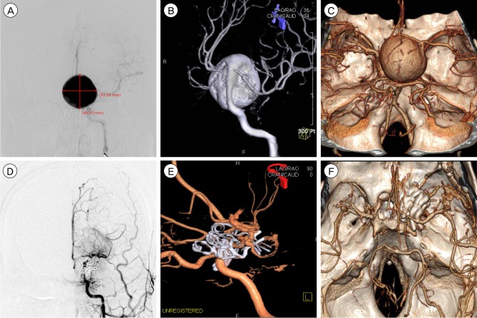
Fig. 1.
Preoperative left digital subtraction angiography (DSA) (A, B) representing a giant left internal carotid artery (ICA) posterior wall aneurysm. (A) Anteroposterior view. (B) Anterooblique view of three-dimensional (3-D) DSA. (C) 3-D computed tomographic (CT) angiography showing a giant sac adhering to all of the surrounding anterior and posterior cerebral arteries on both sides. Postoperative DSA (D, E) representing complete obliteration of the giant aneurysm sac and reconstruction of the ICA using eight different shapes of fenestrated clips. (D) Anteroposterior view (E) Lateral view of 3-D DSA. (F) Postoperative 3-D CT angiography.

