
Fig. 2.
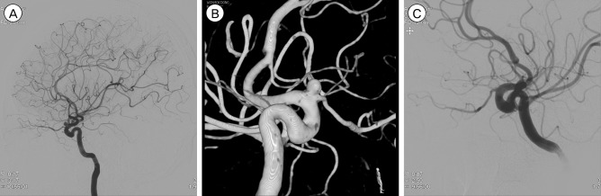
Preoperative left digital subtraction angiography (DSA) (A, B) representing a left ophthalmic artery aneurysm. (A) Lateral view. (B) Lateral view of 3-D DSA. (C) Lateral view. Coil embolization was performed without using a stent. Postoperative left DSA (C) representing complete coil packing of the left ophthalmic artery aneurysm and showing that left ophthalmic artery flow was well preserved.