Fig. 5.

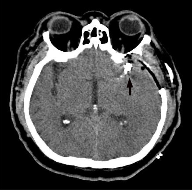
Postoperative computed tomography shows the aneurysmal sac, which was clipped using a clip, and the presence of bony defect because of a prophylactic decompressive craniectomy (black arrow).

Postoperative computed tomography shows the aneurysmal sac, which was clipped using a clip, and the presence of bony defect because of a prophylactic decompressive craniectomy (black arrow).