
Figure 4.
Histological appearance of a small bile duct with inflammatory cells (A) and a small intra-hepatic bile duct with concentric rings of fibrosis (B) in PSC (40× magnification, H&E).
Official websites use .gov
A
.gov website belongs to an official
government organization in the United States.
Secure .gov websites use HTTPS
A lock (
) or https:// means you've safely
connected to the .gov website. Share sensitive
information only on official, secure websites.
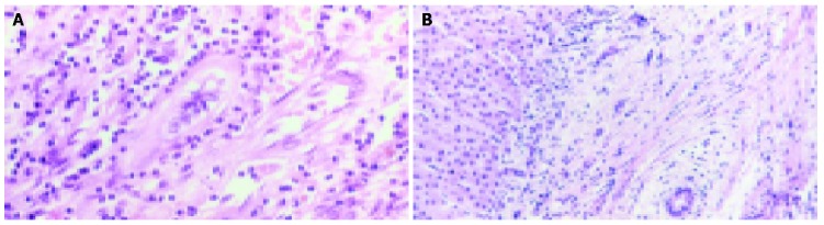
Histological appearance of a small bile duct with inflammatory cells (A) and a small intra-hepatic bile duct with concentric rings of fibrosis (B) in PSC (40× magnification, H&E).