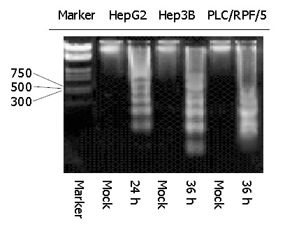
Figure 4.

Kinetics of apoptosis in HepG2 and Hep3B and PLC/RPF/5 cells infected with Ad-TIP30. Twelve, twenty-four or thirty-six hours after infection, analysis of DNA laddering was performed by gel electrophoresis. An overexposed photograph was provided to show the beginning of DNA laddering at 24 h in HepG2 cells and at 36 h in Hep3B and PLC/RPF/5 cells.
