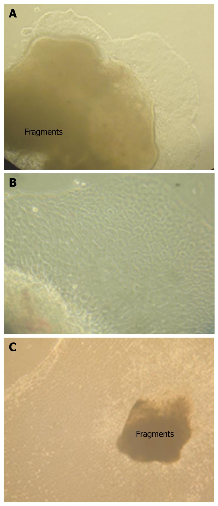
Figure 1.
Isolation of hamster biliary epithelial cells. A: Phase contrast microscopy revealing a small amount of hamster gallbladder epithelial cells growing on the collagen gels 24 h after culture (× 100); B: High magnification of hamster gallbladder epithelial cells demonstrating cuboidal cells around the biliary fragments (× 400); C: Phase contrast microscopy showing widely extended gallbladder epithelial cells on the surface of the gel 7 d after culture (× 40).

