Figure 6.

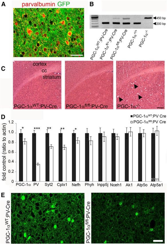
Conditional deletion of PGC-1α in PV-positive interneurons reduces cortical expression of neuron-specific PGC-1α-dependent genes. A, To determine specificity of cre recombinase, PV-Cre mice were crossbred to reporter mice that express GFP after recombination. Somatic GFP expression (green) was restricted to PV (red)-positive cell bodies. Scale bar, 25 μm. B, Efficacy of Cre-mediated recombination was confirmed with qualitative PCR on genomic DNA isolated from whole brains. PGC-1αfl/+:PV-Cre and PGC-1αfl/fl:PV-Cre but not PGC-1αWT:PV-Cre exhibit recombination of the PGC-1α gene (450 bp PGC-1α−/− band). C, Representative photos of hematoxylin and eosin staining demonstrating that PGC-1αfl/fl:PV-Cre mice (6 months) do not develop spongiform lesions in the striatum like PGC-1α−/− mice (P30; arrowheads). Scale bar, 2 mm. cc, Corpus collosum. n = 3 per group. D, qRT-PCR revealed that transcript expression of PGC-1α, PV, Syt2, Cplx1, and Nefh was significantly decreased in PGC-1αfl/fl:PV-Cre compared with PGC-1αWT:PV-Cre cortex. *p < 0.05; **p < 0.005; ***p < 0.0005, one-tailed t tests. E, Protein expression of PV was confirmed to be decreased by immunofluorescence staining (n = 3–4 per group). Scale bar, 50 μm. Numbers per group are indicated on the bar histogram. Data are presented as mean ± SEM.
