Figure 7.
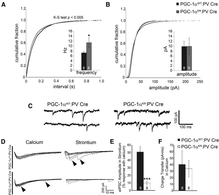
Ablation of PGC-1α in PV-positive interneurons increases asynchronous GABA release in the cortex. Recordings of IPSCs in cortical layer V pyramidal cells were conducted to determine the physiological consequences of PGC-1α deletion. A, The frequency of miniature IPSCs was significantly increased in PGC-1αfl/fl:PV-Cre compared with PGC-1αWT:PV-Cre cortex. B, Miniature IPSC amplitude was not affected. C, Representative traces of spontaneous miniature IPSCs. D, Representative traces showing evoked IPSCs from PGC-1αWT:PV-Cre and PGC-1αfl/fl:PV-Cre mice recorded in normal concentrations of extracellular calcium (left) and after replacing calcium with equimolar concentrations of strontium (right). Traces represent 10 stimulus sweeps from the same cell. Late asynchronous events after strontium application (arrowheads; left) were present in PGC-1αfl/fl:PV-Cre neurons under normal physiological conditions (arrowheads; right). E, Evoked IPSC amplitudes were significantly decreased in PGC-1αfl/fl:PV-Cre compared with PGC-1αWT:PV-Cre mice. F, No changes were observed in charge transfer (evoked IPSC area under the curve), a measure of total GABA release. Numbers per group are indicated on the bar histograms. *p < 0.05; **p < 0.005, one-tailed t tests. Data are presented as mean ± SEM.

