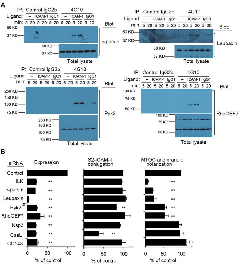
Fig. 3. Signaling Components Required for β2 Integrin-Dependent Granule Polarization.

(A) NK cells treated as in Fig. 2A were tested for the presence of γ-parvin, Leupaxin, Pyk2 and RhoGEF7 in tyrosine-phosphorylated protein complexes. Representative immunoblots of 3 to 5 experiments are shown. (B) mRNA abundance, conjugate formation with S2–ICAM-1 cells, and MTOC and granule polarization, as indicated, of NK cells after silencing of the indicated genes by siRNA. (#Pyk2 protein was monitored by immunoblot.) Data are shown as percent of control siRNA. Conjugation with S2–ICAM-1 cells varied from 32% to 38%, and MTOC and granule polarization varied from 34% to 38% in the controls. Graphs show mean ± SEM from 3 experiments. *p<0.05, **p<0.01.
