
Figure 3. Monosynaptic and Glutamatergic BLA Inputs to the vHPC Are Sufficient to Mediate Changes in Anxiety-Related Behaviors.
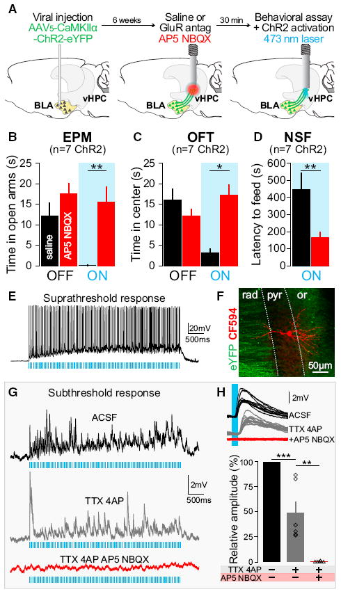
Experiments were performed 6–7 weeks after AAV5-CaMKIIα-ChR2-eYFP injections in the BLA. Means are represented as ±SEM. See also Figures S6–S8. (A) Thirty minutes before behavioral assays and laser stimulation, glutamate receptor antagonists (GluR antag: AP5+NBQX, red) or saline (black) were unilaterally infused locally into the vHPC using the same guide cannula to be used for light delivery via an optical fiber. (B–D) Unilateral AP5+NBQX injection in the vHPC significantly attenuated light-induced increases in anxiety-related behaviors in the EPM, OFT, and NSF relative to saline trials. (B) Intra-vHPC GluR antag microinjection in mice expressing ChR2 in BLA axon terminals blocked the light-induced decrease of open-arm exploration (two-way ANOVA group × light epoch interaction F2,24 = 7.35, p = 0.0032); Bonferroni post hoc analyses showed that mice spent significantly more time in the open arm during light stimulation after intra-vHPC glutamate receptor antagonism (**p < 0.01), but unilateral GluR antag injection did not affect the basal level of open-arm exploration (p > 0.05). (C) After unilateral intra-vHPC glutamate receptor blockade, photoactivation of ChR2-expressing BLA terminals in the vHPC failed to decrease center exploration as seen after saline-treatment trials (two-way ANOVA, F2,24 = 4.17, p = 0.0279, Bonferroni post hoc, *p < 0.05). GluR antag injection did not affect the basal level of open-arm exploration (p > 0.05). (D) GluR antag injection into the vHPC attenuated light-induced increase of latency to feed as seen after saline treatment (one-tailed, paired Student’s t test, n = 7, t = 3.24, **p = 0.0088). (E) Suprathreshold response to photostimulation (20 Hz, 5 ms pulses) of a CA1-vHPC neuron recorded ex vivo in the pyramidal layer (current clamp). (F) Confocal image of a neuron recorded, filled with biocytin and visualized by streptavidin-CF594 reaction (red). Inputs coming from BLA transfected neurons (eYFP). Dotted lines represent the borders of CA1 pyramidal layer (pyr) surrounded by the stratum oriens (or) and stratum radiatum (rad). (G) Subthreshold response of a CA1-vHPC neuron to photostimulation (ACSF, black, 20 Hz, 5 ms pulses). Photoresponse remains after the application of sodium and potassium channel blockers (TTX+4AP, gray) and is abolished by application of GluR antag (TTX+4AP+AP5+NBQX, red). (H) Overlay of ten responses evoked by 5 ms light pulses (ACSF, black), under TTX+4AP perfusion (gray), and after addition of GluR antag (TTX+AP4+NBQX+AP5, red). Histogram population analysis of light-induced EPSP amplitudes confirm that photo-induced EPSP persists after TTX+4AP perfusion (one-way ANOVA, F2,10 = 53.71, ***p < 0.001, Tukey post hoc analysis, ***p < 0.001) and are abolished after application of GluR antag (Tukey post hoc analysis, **p < 0.01).