Figure 2.
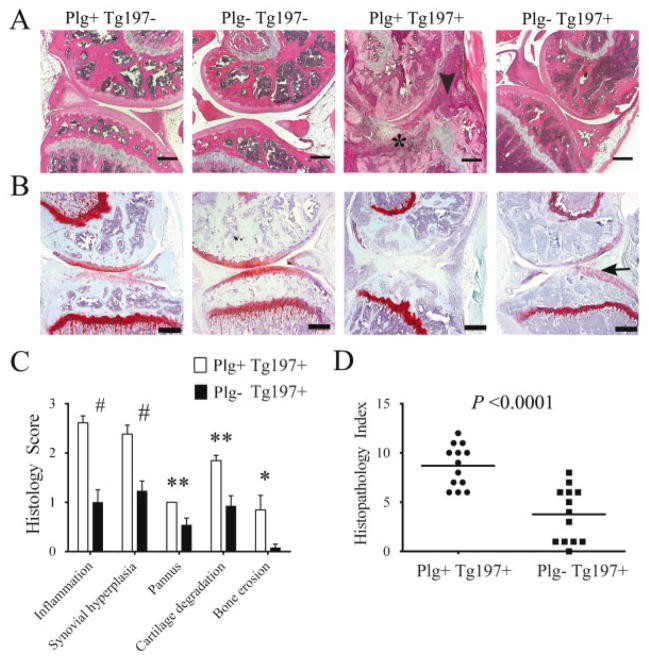
Plasminogen deficiency ameliorates arthritis in the knees of tumor necrosis factor α–transgenic mice. A, Representative hematoxylin and eosin (H&E)–stained knee joint sections from 10-week-old Tg197− and Tg197+ mice with and without plasminogen. Note that widespread joint pathology, including synovial hyperplasia (arrowhead) and pannus (asterisk), was prominent in Plg+/Tg197+ mice, whereas joint architecture was near normal in the Plg−/Tg197+ mice. In the absence of the Tg197 transgene, mouse knee joints were uniformly unremarkable, regardless of the presence or absence of plasminogen. B, Representative Safranin O–stained knee joint sections from 10-week-old Tg197− and Tg197+ mice with and without plasminogen. Note the presence of contiguous red staining indicating intact articular cartilage (arrow) in the Plg−/Tg197+ mice, in contrast to Plg+/Tg197+ mice with extensive articular cartilage destruction. C, Quantitative microscopic analysis of individual disease metrics from H&E-stained knee joint sections from Plg+/Tg197+ mice and Plg−/Tg197+ mice. Bars show the mean ± SEM (n = 13 knees per group). # = P < 0.001; ** = P < 0.01; * = P < 0.05, by Student’s t-test. D, Scatterplot of composite histopathology index analysis of H&E-stained knee joint sections. Symbols represent individual mice; horizontal bars show the median. P values were determined by Mann-Whitney U test. Bars in A and B = 200 μm.

