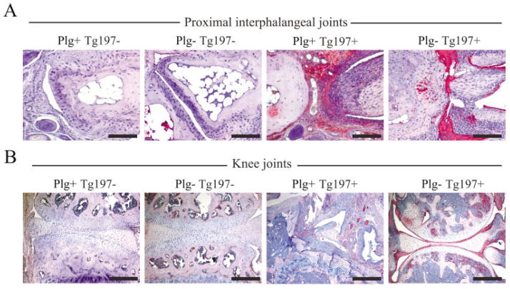
Figure 3.
Fibrin deposition is a consistent histologic feature of joint tissue in plasminogen-deficient mice despite the opposing differences in arthritis disease severity in the paw and knee joints. A, Immunohistochemical staining for fibrin or fibrinogen in representative proximal interphalangeal joint sections isolated from 10-week-old Tg197− and Tg197+ mice with and without plasminogen. Note the robust fibrin deposition (red staining) in the hyperplastic synovium and eroding cartilage surfaces in Plg−/Tg197+ mice. B, Immunohistochemical staining for fibrin deposition in representative knee joint sections isolated from 10-week-old Tg197− and Tg197+ mice with and without plasminogen. Intense fibrin staining was consistently observed in the knee joints of Plg−/Tg197+ mice, particularly along articular surfaces, despite the general lack of microscopic features of arthritis. Not surprisingly, fibrin deposition was also observed within the appreciably more disrupted knee joints of Plg+/Tg197+ mice, often dispersed within the hyperplastic synovial tissue. Bars = 200 μm

