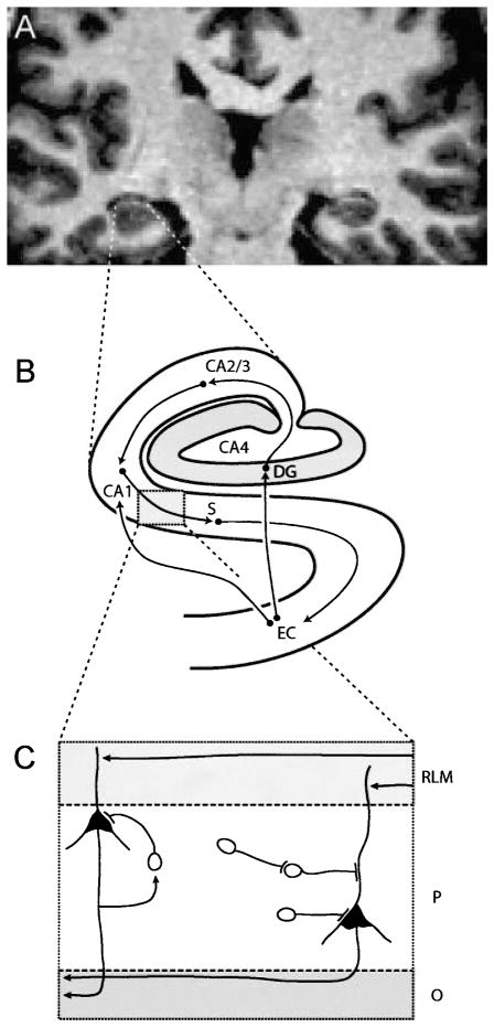
Fig. 2.
Three levels of hippocampal anatomy. A High resolution coronal MRI of the human hippocampus at the level of the hippocampal body. B Schematic diagram of the regions of the human hippocampal formation and their major connections. The entorhinal cortex (EC) sends direct projections and indirect projections via the dentate gyrus (DG) and cornu Ammonis sector CA2/3 to CA1, from which output is relayed via the subiculum (S) back to EC. C The laminar organization of the human hippocampus shown for cornu Ammonis sector CA2/3. Input arrives at dendrites of pyramidal cells in the stratum radiatum/lacunosum/moleculare (RLM), output leaves via fibers in the stratum oriens (O). The two neuronal types in the pyramidal cell layer are the pyramidal shaped principal cells and the nonpyramidal shaped interneuron. At least three types of inhibitory projections can arise from hippocampal interneurons: recurrent inhibition (driven by a collateral of a glutamatergic axon), direct inhibition of a pyramidal cell, and disinhibition of a pyramidal cell (via interneuron-interneuron projection)

