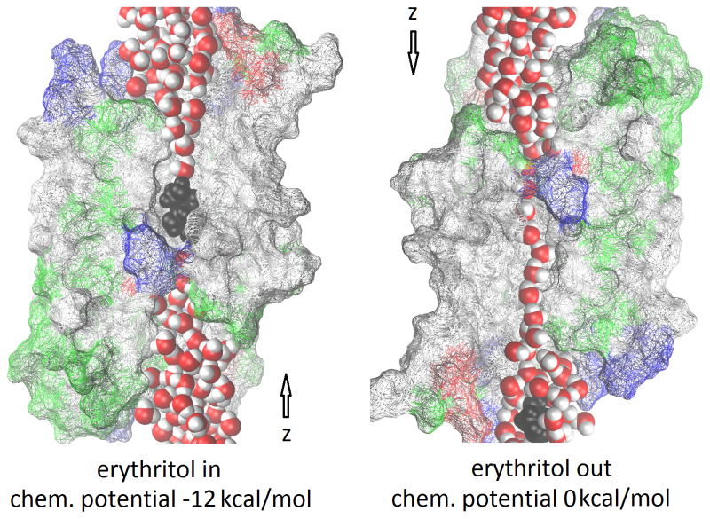
Figure 1.
PfAQP channel occluded (left) and open (right) for water/solute permeation. The luminal residues of the protein are shown in wireframes colored by residue types, the waters in red (oxygen) and white (hydrogen) balls, and the erythritol in black balls (hydrogen, oxygen, and carbon). Not all luminal residues of PfAQP are shown for the purpose of fully exposing the waters and erythritol that line up in single file inside the conducting pore. The z-axis points from the extracellular to the cytoplasmic bulk. Graphics rendered with Virtual Molecular Dynamics. [30]

