
Figure 2.
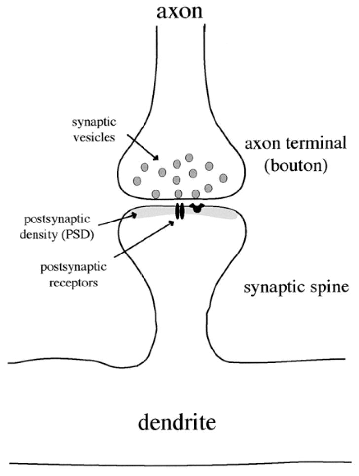
Synapse morphology in the striatum. The majority of neurons in the striatum are medium-size with dendritic spines. Synapses are formed on the dendritic spines as well as on the dendritic shafts (not shown). Synaptic vesicles in the axon terminal release neurotransmitter into the synapse. Neurotransmitters cross the synaptic cleft and interact with receptors in the postsynaptic membrane. The postsynaptic density (PSD) is an electron dense area that contains receptors, ion channels and scaffolding proteins which anchor receptors to the membrane and facilitate signal transduction.