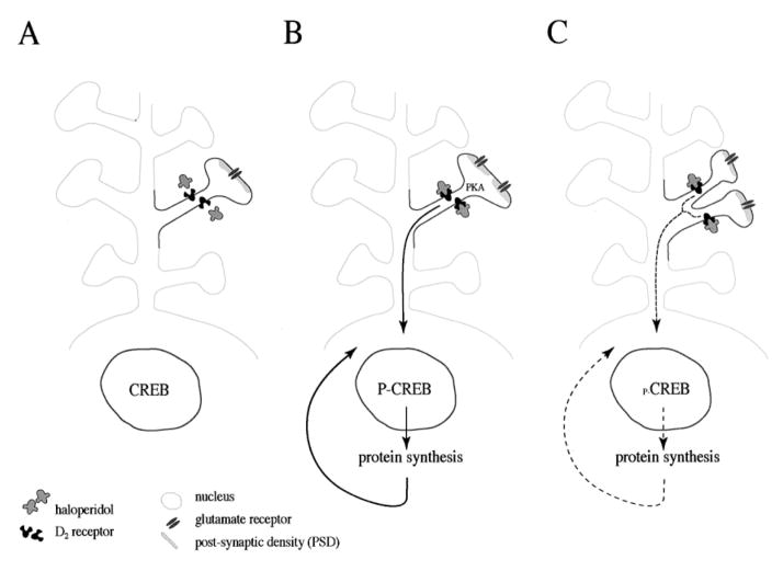
Figure 3.
Gene expression and synaptogenesis by haloperidol. (A) A medium-size spiny neuron in the striatum that expresses D2 receptors contains the transcription factor CREB in the nucleus. (B) Upon interaction of haloperidol and D2 receptors, PKA is activated and proteins are phosphorylated. Phosphorylation of proteins alters their properties and activities and could be the reason for the perforation of synapses (split PSD) (Kerns et al 1992; Meshul and Casey 1989; See et al 1992). PKA also sets a signal transduction cascade in motion that translocates to the nucleus and phosphorylates the transcription factor CREB (Konradi and Heckers 1995). Phosphorylation of CREB at Ser133 (Gonzalez and Montminy 1989) enables the expression of genes and proteins that are under the control of Ca2+ and cyclic AMP second messenger pathways (Montminy 1997). (C) By incorporating newly synthesized proteins, the dendritic spine splits and yields two synapses. Compensatory mechanisms to slow down synaptogenesis will likely develop. Discontinuation of haloperidol administration reverses the process.

