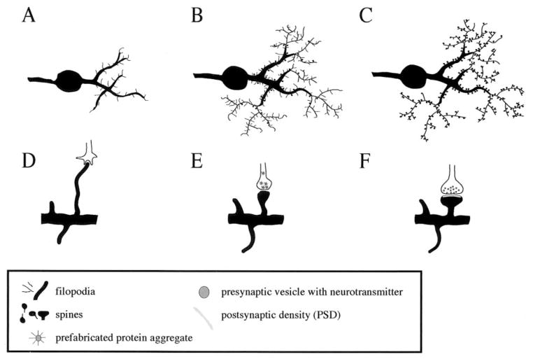
Figure 5.
Synaptogenesis during early neuronal development. (A–C) During early neuronal development, the number of dendritic filopodia decreases while the number of spines increases. (D) An axonal growth cone and a dentritic filopodium make contact and a molecular adhesion process ensues. (E) Prefabricated protein aggregates get trapped at the presynaptic contact zone and enable the axon to release neurotransmitter. (F) The release of neurotransmitter triggers the formation of the PSD.

