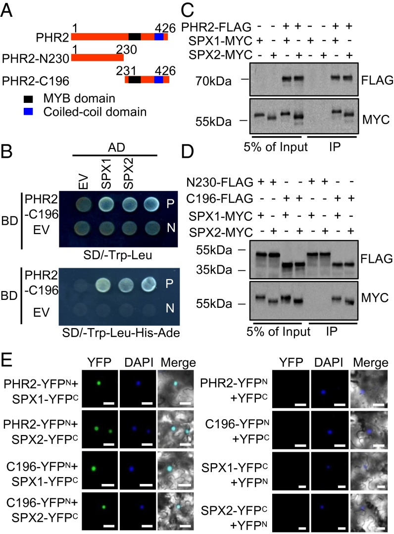
Fig. 1.
SPX1 and SPX2 interact with the PHR2 C terminus. (A) Scheme of full-length PHR2 and deletion derivatives. Numbers above each truncation indicate the PHR2 amino acid coordinates. (B) SPX1 and SPX2 interact with PHR2, as indicated by yeast two-hybrid assays. EV, empty vector; N, negative control; P, positive control. (C) SPX1 and SPX2 interact with PHR2, as indicated by co-IP assays. Protein extracts (Input) were immunoprecipitated with anti-FLAG antibody (IP). Immunoblots were developed with anti-FLAG antibody to detect PHR2 and with anti-MYC to detect SPX1 and SPX2. Molecular mass markers are shown (kDa). (D) SPX1 and SPX2 interact with the PHR2 C terminus, as indicated by co-IP assays. Immunoblots were developed with anti-FLAG and anti-MYC antibodies. Molecular mass markers are shown (kDa). (E) SPX1 and SPX2 interact with PHR2 in the nucleus, as indicated by BiFC analysis. The nucleus was stained with DAPI. (Scale bars: 50 μm.)

