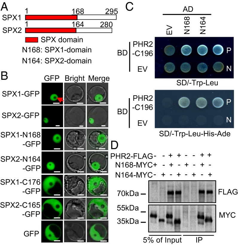
Fig. 2.
Interaction of SPX domains of SPX1 and SPX2 with PHR2. (A) Schematics of SPX1 and SPX2 protein structures. (B) Subcellular localization of full-length and deletion derivatives of SPX1 and SPX2 in rice protoplasts. All truncations were fused with GFP at their C termini under control of the 35S promoter. (Scale bars: 5 μm.) The arrowhead points to the nucleolus. (C) SPX domains of SPX1 and SPX2 interact with PHR2, as indicated by yeast two-hybrid assays. AD, activation domain; BD, binding domain. (D) SPX domains of SPX1 and SPX2 interact with PHR2, as indicated by co-IP assays. Protein extracts (Input) were immunoprecipitated with anti-FLAG antibody (IP) and resolved by SDS/PAGE. The immunoblots shown were developed with anti-FLAG antibody to detect PHR2 and with anti-MYC antibody to detect SPX1-N168 and SPX2-N164. Molecular mass markers are shown (kDa).

