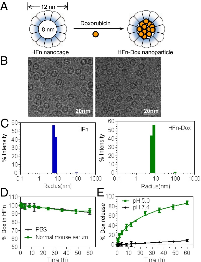
Fig. 1.
Preparation and characterization of HFn-Dox NPs. (A) Schematic depiction of the Dox loading process. (B) Cryo-EM images of HFn nanocages (Left) and HFn-Dox NPs (Right). (C) DLS analysis of HFn nanocages and HFn-Dox NPs. (D) Stability of HFn-Dox NPs in mouse serum at 37 °C over 60 h of incubation (n = 3, bars represent means ± SD). (E) The kinetics of Dox release from HFn-Dox at pH 5.0 and pH 7.0 at 37 °C (n = 3, bars represent means ± SD).

