Fig. 2.
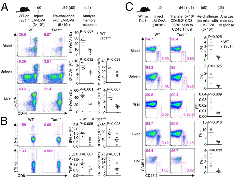
Tsc1 deficiency impairs the recall response of memory CD8+ T cells. (A) WT and Tsc1−/− mice were infected with LM–OVA and rechallenged with LM–OVA 35 d later. The recall response of memory CD8+ T cells was examined at day 4 after secondary infection. Shown are representative flow cytometry plots of CD8+ T cells (Left) and the frequency (Center) and number (Right) of Kb-OVA+ CD8+ T cells. (B) At day 4 after the secondary infection, splenocytes from WT and Tsc1−/− mice were isolated and restimulated with the SIINFEKL peptide for intracellular cytokine staining of IFN-γ and TNF-α. Shown are representative flow cytometry plots of CD8+ T cells (Left) and the frequency (Center) and number (Right) of IFN-γ+ and TNF-α+ CD8+ T cells. (C) Memory CD8+ T cells (CD45.2+) sorted from WT and Tsc1−/− mice at day 41 p.i. were transferred into naïve CD45.1+ recipients. At 24 h, the recipients were challenged with LM–OVA and analyzed 5 d later for CD45.1 and CD45.2 staining of CD8+ T cells (Left) and the frequency of CD45.2+ donor cells (Right). BM, bone marrow; PLN, peripheral lymph nodes. Data are representative of two independent experiments and are presented as the mean ± SEM.

