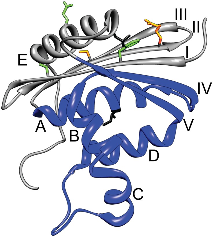
Fig. 1.

Structure of E. coli RNase H (Protein Data Bank 2RN2). Helices are labeled with letters and β-strands with Roman numerals. The region comprising the Icore fragment is colored blue. Residues I25 (on strand II) and I53 (on helix A) are shown in black stick. Other residues shown in stick are involved in the full-length Icore mimics (FL1 contains mutations at I25 and the orange residues, FL2 contains mutations at the orange and green residues).
