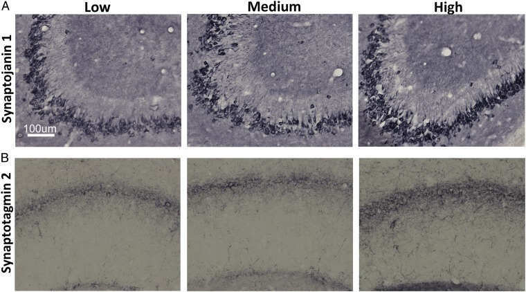
Fig. 4.
High VitD3 enhances expression of synaptic proteins in hippocampal neurons. Representative micrographs of hippocampal sections demonstrate increased immunoreactivity for synaptojanin 1 (A) and synaptotagmin 2 (B) staining in the CA2/3 and CA1 cell layers, respectively, from high-VitD3 animals compared with the low- and medium-VitD3 groups. Synaptojanin 1: optical density of low and medium VitD3 = 0.465 ± 0.0.03, optical density of high VitD3 = 0.54 ± 0.0.02 (P ≤ 0.05). Synaptotagmin 2: optical density of low and medium VitD3 = 0.425 ± 0.0.01, optical density of high VitD3 = 0.442 ± 0.0.008 (P ≤ 0.05).

