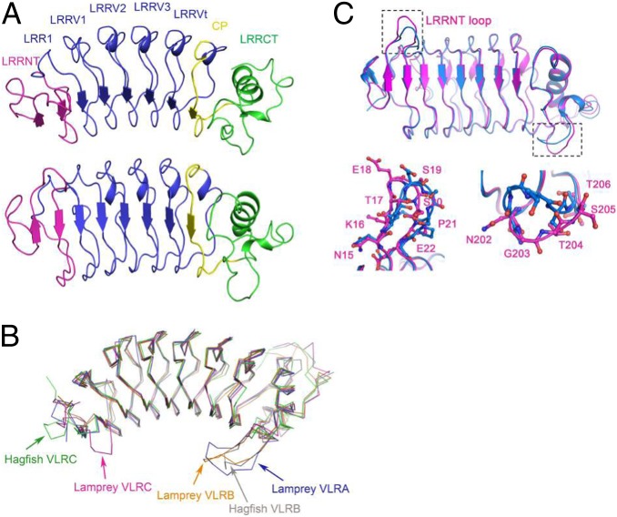
Fig. 3.
Structure of L. planeri VLRC. (A, Upper) Side view of the VLRC.1MP solenoid. LRRNT, magenta; LRR1, LRRV1–3, and LRRVt, blue; CP, yellow; LRRCT, green. (A, Lower) VLRC.1MP rotated 60° from the above orientation to show a front view of the putative antigen-binding concave surface. (B) Comparison of VLRC with VLRA and VLRB. Superposition of VLRC.1MP (magenta) onto lamprey VLRA.R2.1 (PDB accession code 3M18) (blue), lamprey VLRB.aGPA.23 (4K5U) (orange), hagfish VLRB.59 (2O6S) (gray), and hagfish VLRC.29 (2O6Q) (green). Superpositions were carried out by deleting the additional LRRV modules of lamprey VLRA.R2.1 and hagfish VLRC.29 relative to VLRC.1MP. The protruding LRRNT loop of VLRC.1MP is marked with a red arrow. The long LRRCT inserts of VLRA and VLRB are indicated with arrows in corresponding colors. (C) Superposition of VLRC.1MP (magenta) onto Japanese lamprey VLRC (3WO9) (27) (marine), highlighting conformational differences in LRRNT (Lower Left) and LRRCT (Lower Right) loops.

