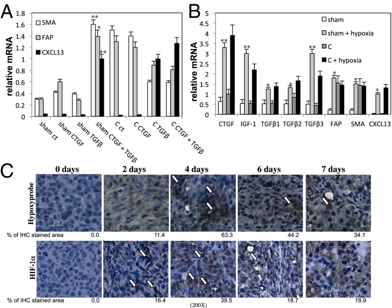
Fig. 3.
Hypoxia caused by androgen ablation induces TGF-β, CTGF, and IGF1 expression. (A) Fibroblasts were purified from Myc-CaP tumors 1 wk after sham operation or castration (C) (n = 10 per group), plated, and stimulated for 24 h with TGF-β1 (10 ng/mL), CTGF (50 ng/mL), or TGF-β1 plus CTGF. RNA was isolated, and expression of the indicated genes was analyzed as described earlier. Results are averages ± SD. (B) Fibroblasts were isolated from Myc-CaP tumors 1 wk after castration (C) or sham operation and incubated either in a standard incubator or a hypoxic chamber (1% O2) for 24 h. Total RNA was isolated, and the indicated mRNAs were quantitated. Results are averages ± SD (C) FVB/N mice bearing Myc-CaP tumors were castrated, and tumors were collected at indicated times after castration. For hypoxia analysis, mice were i.p. injected 90 min before sacrifice with 60 mg/kg of pimonidazole hydrochloride (Hypoxyprobe-1). Tumors were removed, fixed, paraffin-embedded, sectioned, and subjected to IHC with Hypoxyprobe or HIF-1α antibodies. (Magnification: 200×.) The areas occupied by positive cells were determined as described earlier. Arrows indicate hypoxic areas and cells with nuclear HIF-1α.

